- Nucleus -just a scan parameter
-
Nucleus is just a scan parameter
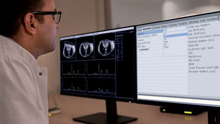
Rather than a complex process, multi-nuclei studies have become a simple protocol that can be “dragged and dropped” into your ExamCard. How much simpler can it be? The nucleus is just a scan parameter like any other sequence parameter. A single ExamCard can be used to run both proton and non-proton imaging and images can be checked on the console before the patient even leaves the room. - Workflow - Proton imaging
-
Workflow does not differ from proton imaging.
We’ve made it easy for your operation, with a seamless integrated workflow for multi-nuclei image acquisition, spectroscopy, reconstruction, and viewing. Reconstruction and viewing of non-proton images or spectra, as well as the process for sending the data to PACS is fully integrated, so workflow does not differ from proton imaging. Easy export of multi-nuclei data is supported for enhanced DICOM, SPAR/SDAT, and XML-REC. - Brain exams, without switching coils
-
Brain exams, without switching coils
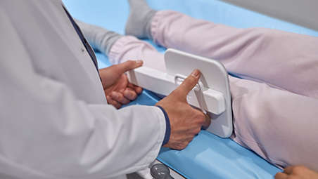
In addition to a seamless user interface, the dual tuned head coils from <b><i><i>RAPID biomedical</i></i></b> enable brain exams, including acquisition of proton and other nuclei, without switching coils. This allows you to schedule your multi-nuclei studies as part of your clinical exam time slots. A full brain study, including both proton (1H) and sodium (23Na) imaging can be completed in 30 minutes¹, all organized in one ExamCard using the same dual tuned head coil. A sodium (23Na) brain exam can be completed in less than 15 minutes². - Improved SNR - Flex coils
-
Improved SNR with transmit-receive flex coils
Transmit-receive flex coils are available for carbon (13C), phosphorus (31P), and sodium (23Na) scans. The ExamCard interface immediately recognizes these multi-nuclei coils. A sodium (23Na) knee exam can be as fast as 15 minutes³. Improved SNR and simplified spectra⁴ can be achieved for phosphorus (31P) and carbon (13C) spectroscopy by combining body coil decoupling, with the transmit-receive surface coils.
Nucleus is just a scan parameter
Nucleus is just a scan parameter
Nucleus is just a scan parameter
Workflow does not differ from proton imaging.
Workflow does not differ from proton imaging.
Workflow does not differ from proton imaging.
Brain exams, without switching coils

Brain exams, without switching coils
Brain exams, without switching coils
Improved SNR with transmit-receive flex coils
Improved SNR with transmit-receive flex coils
Improved SNR with transmit-receive flex coils
- Nucleus -just a scan parameter
- Workflow - Proton imaging
- Brain exams, without switching coils
- Improved SNR - Flex coils
- Nucleus -just a scan parameter
-
Nucleus is just a scan parameter
Rather than a complex process, multi-nuclei studies have become a simple protocol that can be “dragged and dropped” into your ExamCard. How much simpler can it be? The nucleus is just a scan parameter like any other sequence parameter. A single ExamCard can be used to run both proton and non-proton imaging and images can be checked on the console before the patient even leaves the room. - Workflow - Proton imaging
-
Workflow does not differ from proton imaging.
We’ve made it easy for your operation, with a seamless integrated workflow for multi-nuclei image acquisition, spectroscopy, reconstruction, and viewing. Reconstruction and viewing of non-proton images or spectra, as well as the process for sending the data to PACS is fully integrated, so workflow does not differ from proton imaging. Easy export of multi-nuclei data is supported for enhanced DICOM, SPAR/SDAT, and XML-REC. - Brain exams, without switching coils
-
Brain exams, without switching coils
In addition to a seamless user interface, the dual tuned head coils from <b><i><i>RAPID biomedical</i></i></b> enable brain exams, including acquisition of proton and other nuclei, without switching coils. This allows you to schedule your multi-nuclei studies as part of your clinical exam time slots. A full brain study, including both proton (1H) and sodium (23Na) imaging can be completed in 30 minutes¹, all organized in one ExamCard using the same dual tuned head coil. A sodium (23Na) brain exam can be completed in less than 15 minutes². - Improved SNR - Flex coils
-
Improved SNR with transmit-receive flex coils
Transmit-receive flex coils are available for carbon (13C), phosphorus (31P), and sodium (23Na) scans. The ExamCard interface immediately recognizes these multi-nuclei coils. A sodium (23Na) knee exam can be as fast as 15 minutes³. Improved SNR and simplified spectra⁴ can be achieved for phosphorus (31P) and carbon (13C) spectroscopy by combining body coil decoupling, with the transmit-receive surface coils.
Nucleus is just a scan parameter
Nucleus is just a scan parameter
Nucleus is just a scan parameter
Workflow does not differ from proton imaging.
Workflow does not differ from proton imaging.
Workflow does not differ from proton imaging.
Brain exams, without switching coils
Brain exams, without switching coils
Brain exams, without switching coils
Improved SNR with transmit-receive flex coils
Improved SNR with transmit-receive flex coils
Improved SNR with transmit-receive flex coils
Related products
Alternative products
-
Dual tuned head coil
- Perform multi nuclei brain exams in routine scan times
- Acquisition of proton and other nuclei (31P, 13C, 23 Na) without switching coils
- Complete a full brain study, including proton and sodium, in 30 minutes
View product
-
Flex coil C-140
- Transmit-receive C-140 flex coil, with a 14 cm diameter
- Perform carbon (13C) imaging, spectroscopy and research studies, across all anatomies
- Run 13C imaging and spectroscopy directly from the standard user interface
View product
-
Flex coil Na-140
- Transmit-receive Na-140 flex coil, with a 14 cm diameter
- Perform sodium (23Na) imaging, spectroscopy and research studies, across all anatomies
- Run 23Na imaging and spectroscopy directly from the standard user interface
View product
-
Flex coil P-140
- Transmit-receive P-140 flex coil, with a 14 cm diameter
- Perform phosphorus (31P) imaging, spectroscopy and research studies, across all anatomies.
- Run 31P imaging and spectroscopy directly from the standard user interface
View product
-
MR 7700
- XP gradients with high accuracy, power, and endurance
- Facilitate the most demanding research programs
- Supporting confident diagnosis for every patient
View product
-
Ingenia Elition 3.0T X
- A revolutionary breakthrough in diagnostic quality and speed
- Setting new directions for clinical and research 3.0T imaging
- Based on gradient designs with high accuracy, power, and endurance
View product


-
Dual tuned head coil
The dual tuned head coil from RAPID Biomedical allow you to perform brain exams, including acquisition of proton and other nuclei (31P, 13C, 23Na), without switching coils, in routine scan times. A full brain study, including both proton (1H) and sodium (23Na) imaging can be completed in 30 minutes, all organized in one ExamCard, using the same dual tuned head coil. A Sodium (23Na) brain scan can be completed in less than 15 minutes. Workflow does not differ from proton imaging. Multi-nuclei imaging and spectroscopy can be run and reconstructed directly from the standard user interface. The ExamCard interface immediately recognizes the dual tuned head coil. And the nucleus is just a scan parameter like any other sequence parameter. Viewing of multi-nuclei images and spectra, as well as sending data to PACS, is fully integrated. Combined with our multi-nuclei specialist package, the dual tuned head coil allows to explore new imaging pathways by integrating multi-nuclei studies in your day-to-day workflow.
View product
-
Flex coil C-140
The transmit-receive C-140 flex coil, with a 14 cm diameter, allows to perform carbon (13C) imaging, spectroscopy and research studies, across all anatomies. Benefit from improved 13C signal-to-noise ratio (SNR) and simplified 13C spectra, by combining body coil decoupling with this transmit-receive surface coil. Workflow does not differ from proton imaging. 13C imaging and spectroscopy can be run and reconstructed directly from the standard user interface. The ExamCard interface immediately recognizes the C-140 flex coil. And the 13C nucleus is just a scan parameter like any other sequence parameter. Viewing of 13C images and spectra, as well as sending data to PACS, is fully integrated. Combined with our multi-nuclei specialist package, the transmit-receive C-140 flex coil delivers the confidence to explore new imaging pathways and the speed to integrate multi-nuclei studies in your day-to-day workflow.
View product
-
Flex coil Na-140
The transmit-receive Na-140 flex coil, with a 14 cm diameter, allows to perform sodium (23Na) imaging, spectroscopy and research studies, across all anatomies. Benefit from routine scan times and perform a sodium (23Na) knee exam as fast as 15 minutes¹. The sub-millisecond TE acquisition for sodium (23Na) imaging facilitates imaging of short T2-signals. Workflow does not differ from proton imaging. 23Na imaging and spectroscopy can be run and reconstructed directly from the standard user interface. The ExamCard interface immediately recognizes the Na-140 flex coil. And the 23Na nucleus is just a scan parameter like any other sequence parameter. Viewing of 23Na images and spectra, as well as sending data to PACS, is fully integrated. Combined with our multi-nuclei specialist package, the transmit-receive Na-140 flex coil delivers the confidence to explore new imaging pathways and the speed to integrate multi-nuclei studies in your day-to-day workflow.
View product
See all related products -
Flex coil P-140
The transmit-receive P-140 flex coil, with a 14 cm diameter, allows to perform phosphorus (31P) imaging, spectroscopy and research studies, across all anatomies. Benefit from improved 31P signal-to-noise ratio (SNR) and simplified 31P spectra¹. Start to measure the dynamics of muscle metabolism using 31P spectroscopy by visualizing the changes in PCr / Pi-ratio over time. Workflow does not differ from proton imaging. 31P imaging and spectroscopy can be run and reconstructed directly from the standard user interface. The ExamCard interface immediately recognizes the P-140 flex coil. And the 31P nucleus is just a scan parameter like any other sequence parameter. Viewing of 31P images and spectra, as well as sending data to PACS, is fully integrated. Combined with our multi-nuclei specialist package, the transmit-receive P-140 flex coil delivers the confidence to explore new imaging pathways and the speed to integrate multi-nuclei studies in your day-to-day workflow.
View product
-
MR 7700
Experience breakthrough innovation in 3.0T imaging with the unique design of the Philips MR 7700 imaging system, enhanced with XP gradients and artificial intelligence (AI). The system is built to address a pressing need to deliver on the clinical expectations of today, and to facilitate the most demanding research programs. The MR 7700 provides high accuracy, power, and endurance to support confident diagnosis for every patient. It is the system of choice for highest quality diffusion imaging and advanced neuroscience. Extend your scanning capabilities with a fully integrated multi-nuclei imaging and spectroscopy solution to explore new clinical pathways without sacrificing clinical imaging workflow or wide-bore patient comfort. What’s more? The MR 7700 promises a great experience for both users and patients through the ease-of-use features of a well-designed clinical 3.0T scanner together with a no compromise workflow. Now scientists and clinicians alike can schedule without conflict.
View product
-
Ingenia Elition 3.0T X
The Philips Ingenia Elition solution offers cutting-edge MR imaging techniques, while setting new standards for clinical research in 3.0T imaging based on gradient- and RF designs. The Ingenia Elition delivers on superb image quality, and performs MRI exams up to 50% faster¹. Fast overall exam-time is achieved by improving patient handling setup time at the bore with the touchless guided patient setup, combined with accelerations in both 2D- and 3D scanning. Furthermore, the Ingenia Elition offers an immersive audio-visual experience to calm patients and guide them through MR exams.
View product
- 1 Measured from start of first scan to end of last reconstruction. Includes 1H (T2w TSE, T2w FLAIR, SSh DWI, and 3D T1w FFE pre&post) + 23Na (with a voxel size of 4mm isotropic).
- *Caution: Investigational device for imaging with fluorine (19F). Limited by federal (or United States) law to investigational use. Clinical imaging with this nucleus requires usage of a cleared drug. No FDA-cleared drugs are currently available for this nucleus.
- 2 For 4 mm isotropic voxels
- 3 For 3 mm isotropic voxels, slice coverage > 95 mm
- 4 Compared to non-decoupled spectroscopy results