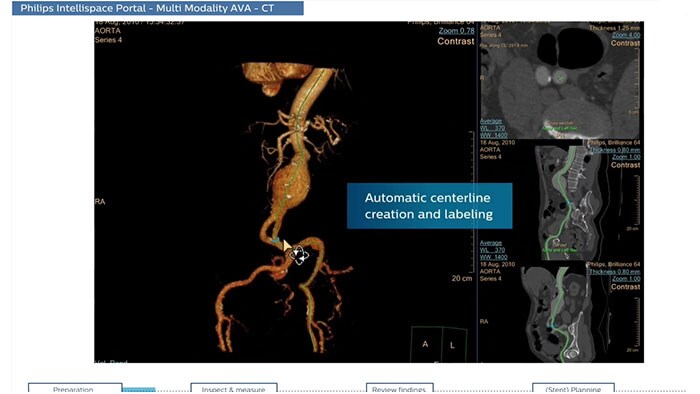
Multi Modality Advanced Vessel Analysis comprehensive vascular planning
The application is designed to examine and quantify different types of vascular lesions from CT and MR, providing modes of inspection, labeling of different vascular lesions, and customizable volume rendering.
Expanding your view of the heart and blood vessels
Take advantage of a comprehensive set of multi-modality advanced visualization vascular applications to examine and quantify vascular lesions. Accelerate workflows with customized views, and enhance workflows for specific findings creation.
One step closer to treatment
Bring advanced diagnostic imaging closer by integrating your Allura / Azurion Interventional Suite with the IntelliSpace Portal, which automatically retrieves patient data from Portal for your scheduled patients.
- Acute Multi-Functional Reivew (AMFR)
-
CT Acute MultiFunctional Review (AMFR)
One application for the assessment of selected anatomies
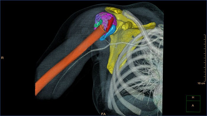
CT Acute MultiFunctional Review (AMFR) provides dedicated tools for findings detection, visualization and assessment of vessels, bones and spine anatomies in 2D and 3D CT images.
Benefits
- Automatic navigation-path for calculation of the spinal cord as well as automatic detection and labeling of spine vertebrae and discs.
- Bones segmentation using an interactive segmentation tool to create a workspace for virtual repositioning of individual bone segments.
- Provides segmentation, editing and measurement tools for vascular analysis.
- Predefined layouts per anatomical area: head, chest, abdomen, spine and extremities.
- Advanced Vessel Analysis (AVA) Stent Planning
-
CT Advanced Vessel Analysis (AVA) Stent Planning
Endovascular stent placement
CT Advanced Vessel Analysis (AVA) Stent Planning includes multiple preset and user-defined options to gain detailed information for use in stent planning.
Benefits
- The application allows you to export customized results to external reporting systems.
- Spectral Advanced Vessel Analysis
-
CT Spectral Advanced Vessel Analysis
IQon Spectral CT Functionality
Benefits
- Bone removal on different energy levels.
- Spectral plots to characterize plaque and stenosis.
- Different energy results comparison.
- Evaluation of the extent of lumen occlusion.
- Spectral Magic Glass on PACS
-
CT Spectral Magic Glass on PACS*
IQon Spectral CT Functionality
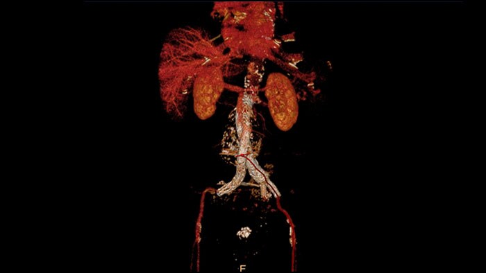
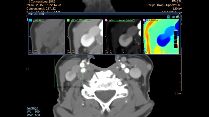
IQon Spectral CT is the only scanner to offer CT Spectral Light Magic Glass and CT Spectral Magic Glass on PACS, helping radiologists review and analyze multiple layers of spectral data at once, including on their PACS.
Benefits
- On-demand simultaneous analysis of multiple spectral results for an Region Of Interest (ROI).
- Integrates into a health system’s current PACS setup for certain PACS vendors.
- Spectral results viewable, during a routine reading.
- Enterprise-wide spectral viewing and analysis allows access to capabilities virtually anywhere in the organization.
* Standard with the CT Spectral option on IntelliSpace Portal.
- Spectral Viewer
-
CT Spectral Viewer
IQon Spectral CT* Functionality
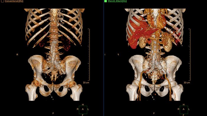
The spectral viewer is optimized for analysis of spectral data sets from the IQon Spectral CT Scanner. Obtain a comprehensive overview of each patient quickly and easily, quantify quickly, and assist in diagnosis. It is designed to accommodate general spectral viewing needs with additional tools to assist in CT images analysis.
Benefits
- Enhances the conventional image by overlaying an iodine map.
- Visualization of virtual non-contrast images.
- Images at different energy levels (40-200 keV).
- Switching to various spectral results can be done through a viewport control.
- Manage presets to create user/site-specific presets.
- Lesion characterization using scatter plots.
- Tissue characterization using attenuation curves.
* IQon CT reconstruction provides a single DICOM entity containing sufficient information for retrospective analysis - Spectral Base Image (SBI). SBI contains all the spectrum of spectral results with no need for additional reconstruction or post-processing. Spectral applications are creating different spectral results from SBI.
- 3D Modeling
-
3D Modeling
Streamlined modeling workflow
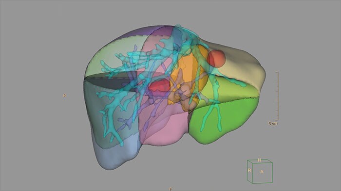
Allows to view volumetric images of anatomical structures, perform segmentation, edit and combine segmented elements (tissues) into a 3D model.
Benefits
- Studies of CT & MR can be used for creating a single 3D model of the same patient. The application provides tools that allow the user to align between the volumes of interest in the images.
- 3D Modeling batches files can be easily exported in standard formats such as STL, with the option to also provide a 3D PDF as an additional means for results sharing with 3D printing or other services* .
- The user may determine the information related to the exported elements of the 3D model such as smoothness and output mesh size.
- Contours can also be exported as RT Structures.
*3D models are not intended for diagnostic use.
- Advanced Vessel Analysis (AVA)
-
Multi Modality Advanced Vessel Analysis (AVA)
Comprehensive vascular analysis planning
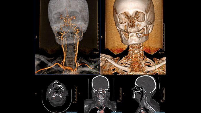
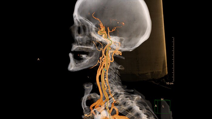
Designed to examine and quantify different types of vascular lesions from CTA and MRA scans. It accommodates different modes of inspection, allows labeling different vascular lesions, and helps navigating through multiple findings.
Demonstrated to reduce the post-processing time by 50% when compared to manual Head & Neck CT angiography (CTA) analysis*.
Benefits
- Ability to choose which Head & Neck Bone Removal method to be used (Standard vs. Smooth).
- Customizable Volume rendering “smoothness” for the 3D Head & Neck vascular structure using a smoothness control.
* Ardley N et al. Efficacy of a new post processing workflow for CTA head and neck. ECR 2013 / C-1760.
- QFlow
-
MR QFlow
Visualizing and quantifying blood flow dynamics
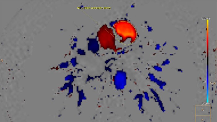
Supports visualization and quantification of blood flow dynamics by assisting in review of MR phase-contrast data, on vascular region of interest segmented manually, or semi-automatically. Qflow analysis is integrated as part of MR Cardiac Suite allowing flow and functional analysis in one suite with combined reporting.
Benefits
- Quantification includes the following parameters: stroke volume, regurgitant fraction, forward and backward flow volumes, flux, stroke distance, mean velocity, maximum velocity, minimum velocity, peak velocity, vessel area, peak pressure gradient, E/A ratio and deceleration time.
- The application supports manual background Correction (BC) to correct for phase (velocity) offset.
- Vascular Processing - DSA (in MMV)
-
XA Vascular Processing - DSA (in MMV)
Contrast arterial structures with surrounding bone and soft tissue to assist in identification of vascular abnormalities
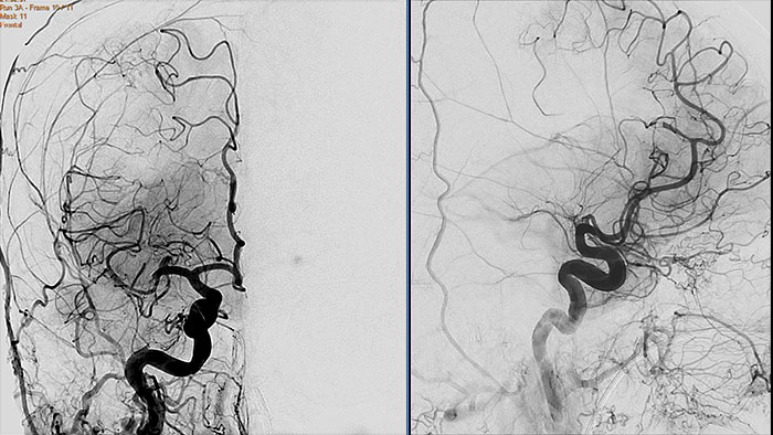
The XA Vascular Processing – DSA (in MMV) expands your workflow by allowing you to read and post-process iXR images virtually anywhere. Obtain images of arteries in various parts of the body using tools to perform standard and run subtractions, pixel shifting, and landmarking. This application also provides post-processing tools to edit and optimize the DSA XA data created in the interventional room.
Benefits
- Single image and ‘Run’ subtraction.
- Pixel Shifting to correct for patient movement during contrast injection and can be performed manually.
- Landmarking helps set a partial subtraction factor.
- Partial subtracted image shows (to a certain degree) the complete image, with the contrast enhanced depending on the subtraction factor.
- Same workflow as in the Philips Allura system.
- Viewing (in MMV)
-
XA Viewing (in MMV)
Comprehensive reviewing tool for multiple modalities, all in a single viewer
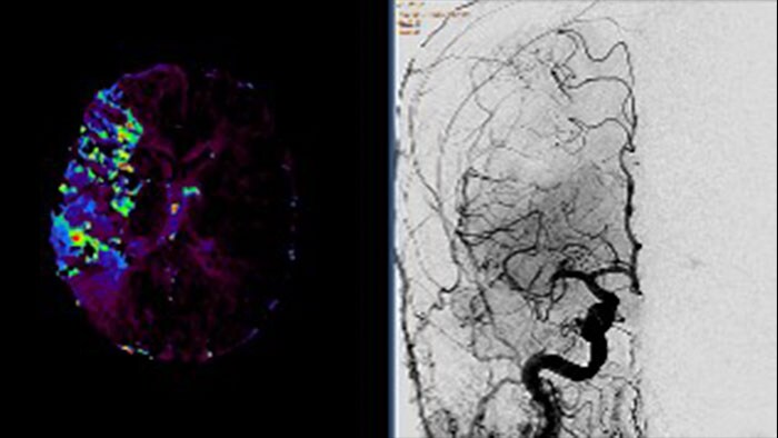
The Multi Modality Viewer (MMV) now supports viewing and post processing of angiography images and series. Review and perform analysis of angiographic imaging alongside other modalities for a comprehensive review of the patient case. Perform vascular processing of images (Digital Subtraction Angiography) – subtraction, pixel shifting and land marking. Include key images into the generic MMV report. Prior to the intervention, relevant diagnostic (MR and or CT) data can be bookmarked and automatically retrieved upon patient selection in the Allura, or the Azurion suites.
Benefits
- Enable a comprehensive review of a patient case across all modalities – CT/MR/NM/US/Angio – in a single environment.
- Advance viewing and post-processing (DSA) of angiographic images and series.
- Annotate and perform basic measurements on images (provided the image is pre-calibrated).
- Automatic retrieval of relevant diagnostic (CT and/or MR) data to support the intervention using iBookmark.
- Reporting also supported; key images can be send to an MMV generic report.
